
The Hidden Players in Brain Function: Glia Take Center Stage
For decades, neurons have been the star of neuroscience research, but a growing body of evidence is shifting the spotlight to a group of non-neuronal cells called glia. These cells, once considered mere support structures for neurons, are now being recognized as essential players in brain health and disease. In fact, the human brain contains as many glial cells as it does neurons, yet our understanding of their organization and functions remains limited.
Scientists at the Salk Institute, led by Terrence Sejnowski, Ph.D., and Shyam Srinivasan, Ph.D., have taken a closer look at glial organization across different brain regions and mammalian species. Their findings, published in PNAS Nexus on October 16, 2025, reveal new insights into how glia shape brain circuit function.
A Comparative Study of Glial Organization
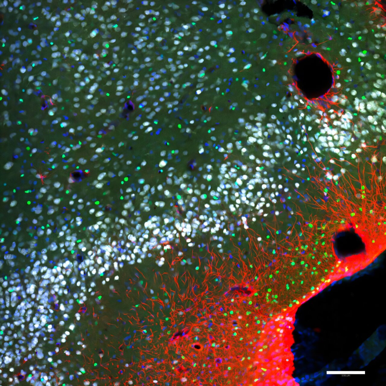
The study focused on four brain regions with distinct functions: the anterior and posterior piriform cortex (involved in smell and emotional processing), the entorhinal cortex (responsible for memory, spatial navigation, and time perception), and the cerebellum (critical for movement, coordination, and learning). By analyzing available samples and datasets from human, rodent, and other mammalian tissues, the researchers were able to compare glial density and the proportion of each cell type—microglia, oligodendrocytes, and astrocytes.
Using techniques such as histology and single-cell RNA sequencing, the team found that both glial density and the relative proportions of these cell types varied significantly across the four brain regions. This suggests that glial organization is not uniform throughout the brain but rather depends on the specific circuitry of each region.
Evolutionary Consistency and Regional Specificity
One of the most significant findings was that these regional differences in glial organization were conserved across mammals. Additionally, the ratio of glia to neurons scaled consistently across species. These results highlight a strong, evolutionarily conserved relationship between glia and neurons, which is specific to each brain region.
For many years, glia were thought to be passive support cells, primarily responsible for maintaining the health of neurons. Earlier studies on the mammalian neocortex suggested that glial organization was relatively uniform. However, this new research challenges that view by showing that each brain region has a unique glial structure that supports its distinct circuitry.
Glia as Markers of Regional Identity
The study also suggests that glia may serve as better markers of regional identity than neurons, especially outside the neocortex. This could have important implications for understanding how different parts of the brain function and how they might be affected in neurological disorders.
Implications for Future Research
The findings from the Salk Institute study underscore the need for future research to explore the role of glia as much as neurons in contributing to brain dysfunction and neurodegenerative diseases such as Alzheimer’s. As scientists continue to unravel the complex interactions between neurons and glia, it becomes increasingly clear that a full understanding of brain function requires examining both.
Other contributors to the study include Antonio Pinto-Duarte and Katharine Bogue of Salk.
Further Reading
For more information, you can refer to the following publication:
Antonio Pinto-Duarte et al, Conservation of glial density and cell-type ratios within a brain region across mammals, PNAS Nexus (2025). DOI: 10.1093/pnasnexus/pgaf314
This research opens up exciting new avenues for studying the brain and could lead to breakthroughs in treating neurological conditions that have long been poorly understood.
Post a Comment